Introduction
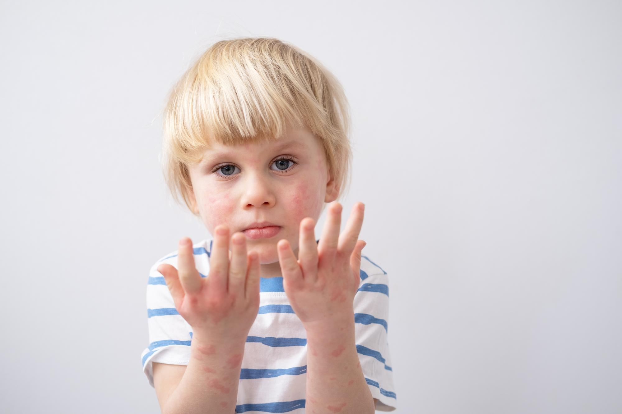
Rashes are frequently reported in pediatrics, yet particular rash patterns can hint at underlying rheumatologic conditions. Outpatient pediatricians, especially general practitioners, must recognize these unique rashes to diagnose issues such as juvenile lupus, dermatomyositis, systemic juvenile idiopathic arthritis, vasculitides, and others like pernio. Prompt identification and precise differential diagnosis are crucial for timely referrals and management. This summary highlights the practical elements of diagnosing and distinguishing rashes associated with pediatric rheumatologic diseases, focusing on clinical utility rather than pathophysiology, serving as a primary resource for clinicians.
When assessing a child’s rash for possible rheumatologic causes, keep these key points in mind:
Rash Morphology, Distribution: Identify the rash type—macular, papular, urticarial (like hives), purpuric (non-blanching), scaly, or nodular—and its location (e.g., face, trunk, extensor surfaces, palms/soles). Some diseases exhibit specific distributions, such as cheeks in lupus, knuckles in dermatomyositis, and legs in vasculitis).
Timing and Triggers: Assess whether the rash is transient or persistent, observing any patterns. For instance, an evanescent rash might correspond with fever spikes in systemic JIA or be provoked by cold exposure in pernio. Additionally, note if the rash intensifies with sunlight, as photosensitivity could indicate lupus or dermatomyositis.
Associated Symptoms: Conducting a comprehensive review of symptoms is essential. A fever pattern characterized by quotidian high fevers may indicate systemic JIA. Look for signs such as arthritis or arthralgia, which could suggest rheumatic conditions like lupus, JIA, vasculitis, muscle weakness (indicative of dermatomyositis), oral or genital ulcers (associated with lupus or Behçet disease), mucositis or conjunctivitis (linked to Kawasaki disease), and Raynaud’s phenomenon (potentially pointing to juvenile scleroderma or lupus). These details can help narrow down the diagnosis differential.
Systemic Findings & Labs: Assessing growth parameters, vital signs, and specific lab tests can provide valuable insights. For instance, cytopenias or elevated ANA titers indicate lupus; increased muscle enzymes (CK, aldolase) suggest dermatomyositis; elevated inflammatory markers alongside anemia and very high ferritin indicate systemic JIA; a normal platelet count with purpura points towards vasculitis (such as IgA vasculitis) instead of thrombocytopenia. While basic screening labs (CBC, ANA, ESR/CRP, UA) can assist in the evaluation, clinical suspicion must lead the process of interpretation.
Approach to Evaluating a Pediatric Rash for Rheumatologic Etiology
Because rashes in rheumatologic diseases are diverse, a systematic approach is helpful. The following algorithm outlines a stepwise evaluation:
- Determine if the rash is blanching or non-blanching. Non-blanching purpuric rashes indicate hemorrhage into the skin:
- When petechiae or purpura are found, checking the platelet count and coagulation is crucial. First, it’s essential to rule out thrombocytopenic purpura, like ITP, or coagulation disorders. If platelet levels are normal and the purpura are palpable, small-vessel vasculitis, such as IgA vasculitis (Henoch–Schönlein purpura), is probable. The appearance of purpura, especially on the lower limbs and buttocks, combined with arthritis and abdominal pain, strongly indicates IgA vasculitis. In contrast, widespread purpura that occurs with fever and a severely ill appearance raises concerns about meningococcemia or other severe infections (emergency).
- If the rash is blanching (erythematous), assess its morphology and context:
- Localized facial erythema is characterized by a red rash localized to the cheeks (malar region), often a sign of acute cutaneous lupus, especially if the nasolabial folds remain unharmed. This malar rash, commonly called the “butterfly” rash, tends to be sensitive to light and may last from several hours to a few days during flare-ups. It is critical to assess for other lupus symptoms, such as oral ulcers, arthritis, and kidney-related signs, and conduct an ANA test. Differential diagnoses include the fifth disease (erythema infectiosum), which manifests as a “slapped-cheek” rash mainly in younger children and often appears with a lacy body rash. Disorders like rosacea or seborrheic dermatitis can cause facial redness but are not limited to the malar region and have different chronic patterns. Unilateral cheek redness accompanied by fever may indicate erysipelas or cellulitis, which are generally warm and tender. Dermatomyositis can also lead to facial erythema, often showing as a violaceous periorbital (heliotrope) rash (see the section) below).
- Violaceous periorbital and hand rash: Juvenile dermatomyositis (JDM) presents with a heliotrope rash, characterized by a purple discoloration of the eyelids, alongside Gottron papules, which are violaceous lesions found on the extensor joints of the hands, such as the knuckles, elbows, and knees. When a child displays these distinctive rashes with muscle weakness, JDM is likely the primary diagnosis, as skin manifestations generally occur before muscle symptoms. Differentials: Though lupus may occasionally lead to periorbital swelling or rashes, the characteristic heliotrope color, and Gottron papules are exclusive to dermatomyositis. Additionally, periorbital allergic reactions or infections, such as orbital cellulitis, can produce swelling or discoloration but do not form the distinctive papules joints.
- Consider systemic juvenile idiopathic arthritis (sJIA) when a patient displays a generalized maculopapular or urticarial-like rash with fever, especially if the rash is transient and aligns with daily high fevers. sJIA-related rashes typically present as distinct salmon-pink macules or slightly raised lesions, mainly found on the trunk and proximal limbs during fever spikes, and they fade as the fever resolves. The rash usually does not itch, though it can be induced by scratching (known as the Koebner phenomenon). Monitor for a daily fever pattern and signs of arthritis, hepatosplenomegaly, or lymphadenopathy to further indicate sJIA. Consider differential diagnoses like viral infections (e.g., adenovirus or EBV), which may cause fevers with rashes, but typically, viral exanthems last longer or are more widespread and don’t necessarily coincide with fever spikes. Drug reactions such as serum sickness can also result in urticarial or maculopapular rashes. Reviewing the medication history is essential in history. Autoinflammatory syndromes like TRAPS or CAPS can lead to recurrent fevers with rashes, although rarer; for instance, TNF-receptor-associated periodic syndrome might show a migratory red rash during fever episodes. If fever and rash last more than five days in a young child, consider Kawasaki disease or MIS-C (Multisystem Inflammatory Syndrome in Children) COVID-19).
- Polymorphous rash with mucosal involvement: A child under 5 with a fever lasting 5 days or longer and showing a polymorphous rash might be experiencing Kawasaki disease. This type of rash usually affects the trunk, limbs, and frequently the diaper area, often peeling during healing. Other diagnostic indicators include red eyes, cracked red lips, a “strawberry tongue,” swollen red hands or feet (which may later peel), and a unilateral cervical lymph node larger than 1.5 cm. Kawasaki disease is a vasculitis that requires prompt treatment to prevent coronary aneurysms. Differential diagnoses include viral exanthems (measles, adenovirus, enterovirus), which can mimic the Kawasaki rash but typically have distinctive features such as cough, runny nose, and conjunctivitis in measles and a unique rash progression from head to toe. Scarlet fever (a streptococcal infection) results in a widespread sandpapery scarlatiniform rash and a strawberry tongue. Still, this rash typically appears before the fever, and streptococcus can be confirmed via throat culture. Staphylococcal scalded skin syndrome and toxic shock syndrome may present with fever along with rash and peeling, yet children with these syndromes generally appear more ill. Stevens-Johnson syndrome (erythema multiforme major) presents mucositis and rash, usually characterized by target lesions or bullae, along with a history of medication exposure. Since 2020, Multisystem Inflammatory Syndrome in Children (MIS-C) linked to COVID-19 has been included in the differential diagnosis; it often features a persistent fever, a rash (which may be polymorphic or purpuric), and multi-organ involvement, frequently overlapping with incomplete Kawasaki disease.
- Chronic scaly (“psoriasiform”) plaques: When a rash features erythematous or violaceous scaly plaques, particularly on extensor surfaces like the knees, elbows, scalp, or behind the ears, psoriasis should be considered. In children with arthritis—especially those showing asymmetric or distal interphalangeal joint arthritis or dactylitis in fingers and toes—psoriasis could indicate juvenile psoriatic arthritis, a subtype of JIA. Classic psoriasis plaques are well-defined reddish or purplish patches adorned with white or silvery scales. Guttate psoriasis, marked by small drop-like lesions with fine scaling, can follow a streptococcal infection in children. Nail changes such as pitting or onycholysis are commonly associated with psoriasis. Differential diagnoses: Atopic dermatitis (eczema) can also cause a chronic itchy rash; however, eczema in school-aged children typically affects flexural areas such as the antecubital and popliteal fossae, appearing less well-defined and usually without the thick silvery scales characteristic of psoriasis. Tinea corporis (a dermatophyte infection) may present as ring-shaped scaly plaques with central clearing and a raised border; confirming fungi’s presence can be confirmed with a KOH scraping. Pityriasis rosea leads to multiple oval scaly patches on the trunk, is self-limiting, and is not linked to arthritis.
- Tender subcutaneous nodules, especially on the shins, suggest erythema nodosum, a reactive inflammatory disorder affecting fat tissue rather than a specific rheumatic disease. In children, this condition may arise from infections (like streptococcal or tuberculosis), medications, or underlying inflammatory diseases such as Behçet disease or inflammatory bowel disease. Although Behçet disease is uncommon in children, it can manifest with lesions that resemble erythema nodosum and an acneiform rash, distinguished by its defining symptoms: recurrent and painful oral and genital ulcers, along with ocular inflammation. For differential diagnoses, bruises or trauma might be mistaken for nodules; cellulitis can cause painful, red subcutaneous areas but is typically unilateral and warmer.
- Livedo reticularis/racemosa: A net-like purple skin discoloration can occur in vasculitic or prothrombotic disorders. In children, livedo reticularis is often a harmless response to cold; however, if it becomes persistent (racemosa) and is accompanied by ulcers or neurological signs, it may indicate antiphospholipid syndrome or medium-vessel vasculitis, such as polyarteritis nodosa. While these conditions are uncommon, recognizing them is crucial; a rheumatology referral is recommended and warranted.
- Evaluate for other potential causes or triggers: Some rheumatologic rashes may arise from various disorders. For example, pernio (chilblains) usually results from cold exposure, but it can also indicate lupus (chilblain lupus) or other autoimmune diseases. If a child shows persistent or severe pernio lesions or develops pernio in milder climates, conduct an ANA test for lupus and assess for systemic symptoms. Similarly, if a rash that resembles psoriasis occurs alongside arthritis, consider the possibility of psoriatic arthritis. If recurrent mouth sores appear with a rash, keep Behçet’s disease in mind, among other options. It’s also important to review medications to rule out drug rashes and infection exposures.
- Conduct focused investigations or referrals: Based on the suspected diagnosis, obtain suitable confirmatory tests. For instance, if lupus is suspected, test for ANA, dsDNA, complement levels, and perform a urinalysis; for JDM, check muscle enzymes, consider an MRI, or refer for nailfold capillaroscopy and potentially a muscle biopsy; for sJIA, assess ferritin, IL-18 (if available), and monitor for macrophage activation syndrome; for HSP, perform a urinalysis to evaluate kidney involvement, and consider a skin biopsy if the presentation is atypical; for suspected Kawasaki disease, conduct echocardiography and check inflammatory markers. Early involvement of a pediatric rheumatologist or dermatologist can be crucial in uncertain cases. The algorithm’s summarized approach helps ensure both common and serious conditions are considered. Below, we elaborate on the main pediatric rheumatologic conditions and their related rashes, followed by summary tables and a downloadable resource algorithm.
Rashes in Specific Pediatric Rheumatologic Diseases
Juvenile Systemic Lupus Erythematosus (JSLE) – Malar and Other Lupus Rashes
Skin symptoms are prevalent in juvenile-onset SLE, affecting around 60-85% of patients. The hallmark rash is the malar (butterfly) rash, characterized by redness on the cheeks and nose with sparing of the nasolabial folds. This rash often appears or worsens following sun exposure (photosensitivity) and may be linked to disease activity. Unlike a typical sunburn, the malar rash in lupus is a persistent red or purplish flush that can last several days without causing pain. Discoid lupus lesions, which are disc-shaped and scaly, can present as either hypopigmented or hyperpigmented plaques that may lead to scarring. Although these are less common in children, they can occur, particularly in chronic cutaneous lupus. Children with SLE may also present with generalized acute rashes (for example, a morbilliform rash on the arms and trunk) during flare-ups, in addition to oral or nasal ulcers and hair loss being significant indicators.
Important diagnostic indicators are photosensitive malar rash, oral ulcers, arthritis, renal symptoms (hematuria/proteinuria), and cytopenias. A malar rash in a female adolescent with these features strongly indicates lupus. A timely referral is recommended, as JSLE typically needs swift treatment to avoid organ damage damage.
Juvenile Dermatomyositis (JDM) – Heliotrope and Gottron’s Papules
JDM is an idiopathic inflammatory myopathy characterized by hallmark skin findings. The heliotrope rash is a pathognomonic indication: a violaceous (purple-red) discoloration of the eyelids, often accompanied by periorbital edema. Accompanying this are Gottron papules, which are violaceous papules or plaques on the extensor surfaces of joints, classically found on the dorsal hands over the knuckles (MCP/PIP joints) and on the elbows and knees. Over time, these skin lesions may develop a scaly or atrophic quality. Other photosensitive rashes associated with JDM include the “shawl sign” (a reddish rash over the shoulders, upper back, and V of the neck/chest) and malar erythema; essentially, sun-exposed areas may exhibit a rash. Nailfold capillary changes (dilated capillary loops or dropout) are another telltale sign, observable with a magnifier or dermatoscope. Skin signs often precede muscle weakness in JDM, so recognizing the rash can lead to early diagnosis before significant muscle damage occurs. Children with JDM typically develop symmetrical proximal muscle weakness (difficulty climbing stairs, getting off the floor, lifting arms) several days to weeks after the rash onset. Calcinosis cutis (calcium deposits in the skin) may occur in chronic cases.
Key diagnostic indicators include heliotrope eyelid rash, Gottron papules, and proximal muscle weakness. Increased muscle enzyme levels (CK, AST, LDH) further affirm the diagnosis. JDM is a critical condition that necessitates immunosuppressive treatment; therefore, if suspected, an urgent referral is indicated.
Systemic Juvenile Idiopathic Arthritis (Still’s Disease) – Evanescent Salmon Pink Rash
Systemic JIA, often referred to as Still’s disease in the context of systemic juvenile arthritis, stands out among the subtypes of JIA due to its prominent fever and rash. The rash associated with systemic JIA is characteristically salmon-pink macules, typically measuring between 2 and 10 mm in diameter, with a slight elevation (maculopapular), primarily appearing on the trunk and proximal extremities. It usually manifests in the evening or at night, coinciding with a peak in daily fever, then fades as the fever subsides, only to reappear with the next fever spike. These lesions are generally non-itchy and may go unnoticed if the child is only examined when they are afebrile. Rubbing or scratching the skin can trigger new lesions, known as the Koebner phenomenon, which may present as linear streaks after scratching. In addition to the rash, systemic JIA is a systemic autoinflammatory disease; children typically endure high, daily (“quotidian”) fevers for a minimum of two weeks, along with other symptoms such as generalized lymphadenopathy, hepatosplenomegaly, and arthritis, which might not be evident at the onset but can develop later in the course of the disease course).
Key diagnostic indicators include persistent fevers lasting over two weeks, a transient salmon-colored rash accompanying the fevers, joint pain or arthritis, and lab results showing systemic inflammation (elevated ESR/CRP, significantly high ferritin, and neutrophil leukocytosis). It is crucial to rule out any infectious causes. The identification of the unique rash aids in differentiating systemic juvenile idiopathic arthritis (sJIA) from other origins of extended fever in children. Early consultation with a rheumatologist is advised for accurate diagnosis and management, which frequently comprises using IL-1 or IL-6 inhibitors sJIA).
IgA Vasculitis (Henoch–Schönlein Purpura, HSP) – Palpable Purpura
IgA vasculitis (IgAV), previously known as Henoch–Schönlein purpura, is children’s most prevalent form of vasculitis. A rash is present in 95% of cases. This rash starts as erythematous maculopapules and evolves into palpable purpura—small, raised purple spots—due to leukocytoclastic vasculitis affecting the skin. Typically, the rash appears symmetrically and is localized to the lower extremities and buttocks (areas affected by gravity). Initially, the lesions may resemble red or pink spots but change into purpuric lesions that do not blanch. Bruising may also occur; bullae or necrotic ulcers can form within the rash in severe instances. While the rash can extend up to the thighs and arms, it is rare for IgA vasculitis purpura to appear on a child’s face or upper trunk. Other essential characteristics of IgA vasculitis include arthralgia or arthritis (often transient and affecting the ankles or knees), abdominal pain (due to GI vasculitis leading to intestinal angina or even bleeding/intussusception), and renal involvement (IgA nephropathy presenting as hematuria or proteinuria). The combination of palpable purpura, abdominal pain, and arthritis in a child is usually diagnostic.
Key diagnostic indicators include palpable purpura on a child’s buttocks or legs, a normal platelet count, and any combination of abdominal pain, arthritis, or hematuria, which strongly suggests IgA vasculitis. Management typically involves supportive care, such as analgesics and hydration; however, it is important to monitor urine for potential kidney involvement over the course of weeks to months. If there is considerable renal involvement or severe gastrointestinal complications, specialty care becomes necessary.
Kawasaki Disease – Polymorphous Exanthem in a Febrile Infant/Toddler
Kawasaki disease (KD) is an acute childhood vasculitis primarily affecting children under 5 years old. One of its hallmark symptoms is the rash, which lacks a singular appearance, leading to its description as “polymorphous.” It can manifest as a generalized maculopapular rash that might resemble a viral rash or lesions similar to erythema multiforme or, occasionally, a fine papular (scarlatiniform) rash. Typically, the rash emerges within the first five days after fever onset. It usually affects the trunk and extremities, with common involvement of the groin and perineum, where peeling can serve as an early indicator. The rash generally does not itch. Key diagnostic features of Kawasaki disease include a persistent fever lasting at least five days, bilateral non-purulent conjunctivitis, changes in oral mucosa (like red, cracked lips and strawberry tongue), alterations in peripheral extremities (such as erythema or swelling of hands and feet, followed by desquamation of fingers and toes), and cervical lymphadenopathy. While not all symptoms appear at once, having four of the five features and fever indicates classic KD. Skin desquamation, particularly around the nails of fingers and toes, typically develops during the subacute phase (2–3 weeks after onset); thus, initial diagnosis often focuses on the rash rather than this peeling.
Key diagnostic indicators: A prolonged fever lasting more than 5 days in a child aged 1 to 5, accompanied by a non-specific erythematous rash, conjunctivitis, oral changes, swollen hands and feet, and enlarged neck lymph nodes, strongly indicates Kawasaki disease. Early identification is vital, as prompt IVIG treatment significantly lowers the chances of developing coronary artery aneurysms. If Kawasaki disease is suspected, arranging an echocardiogram and initiating IVIG and aspirin treatment according to guidelines is essential. In cases where not all criteria are met, laboratory markers such as high CRP, elevated ESR, thrombocytosis, increased liver enzymes, and sterile pyuria, along with echocardiogram results, assist in confirming the diagnosis and deciding whether to proceed with treatment for Kawasaki disease.
Juvenile Psoriatic Arthritis – Psoriatic Rash
Juvenile psoriatic arthritis (JPsA) is a specific type of juvenile idiopathic arthritis (JIA) characterized by the presence of both arthritis and psoriasis (or a significant family history of psoriasis along with arthritis showing certain features). In children, the psoriasis rash can manifest in various forms, with plaque psoriasis being the most common: distinct red, pink, or purplish plaques topped with a white, silvery scale. Typical locations for these plaques include the extensor surfaces of elbows and knees, the scalp, and the back of the ears. Some children may instead exhibit guttate psoriasis, which presents as numerous small drop-like scaly spots across the trunk, often following a streptococcal throat infection. Psoriasis can also occur in skin folds (inverse psoriasis) or solely impact the nails (appearing as pitting, ridging, and oil-drop discoloration). In JPsA, arthritis may affect the finger joints (distal interphalangeal joints) and typically involves dactylitis, where a whole finger or toe swells up like a sausage. Nail pitting is a crucial indicator in pediatric arthritis, even when the skin rash is mild or has not yet developed. Approximately one-third of children with psoriasis will develop arthritis; in some cases, the arthritis can precede the skin rash by several months longer.
Key diagnostic indicators of juvenile psoriatic arthritis include chronic scaly plaques on the extensor surfaces, nail pitting, and dactylitis in affected children. Patients with this condition may also show a positive ANA, which can be present in up to 50% of early-onset cases, and they are at risk for asymptomatic uveitis, as seen in other types of juvenile idiopathic arthritis (JIA). Treatment involves collaboration between dermatology and rheumatology. Approaches may include topical therapies, methotrexate, or biologics to manage both skin and joint issues inflammation effectively).
Localized Scleroderma (Morphea) and Systemic Sclerosis
Scleroderma encompasses autoimmune diseases marked by skin hardening or sclerosis. In children, localized scleroderma, known as morphea, is more prevalent than systemic sclerosis. Morphea presents as one or multiple patches of skin change: initial lesions may appear as erythematous or violaceous patches or plaques that eventually become hard and bound down, exhibiting a whitish or brownish center surrounded by a violaceous border. Over time, these patches may resemble scar tissue. Morphea lesions can take on an oval or linear form. A type of localized scleroderma, linear scleroderma, manifests as a linear band of sclerosis, often on a limb, potentially leading to growth impairment of that limb or on the forehead, forming a line known as “en coup de sabre.” Unlike typical rashes characterized by widespread inflammation, these manifestations signify localized skin autoimmune phenomena. Surface alterations may include alopecia or pigment changes in the affected area. Morphea generally lacks systemic symptoms, remaining confined to the skin and occasionally affecting underlying tissue.
Juvenile systemic sclerosis (JSSc) is infrequent, yet pediatricians should be alert to the Raynaud phenomenon, which involves episodic finger blanching and cyanosis in cold conditions, as it may signify onset. Children with early systemic sclerosis may display puffy, swollen fingers that eventually become hardened and shiny (a condition called sclerodactyly), accompanied by generalized skin tightness. They may develop areas of hyperpigmentation or hypopigmentation that feel stiff. In contrast to morphea, systemic sclerosis impacts internal organs, including the GI tract and lungs, and is often associated with positive systemic antibodies such as anti-centromere antibodies anti-topoisomerase).
Important diagnostic indicators include localized scleroderma, which appears as solitary or a few patches of skin that become hardened, shiny, hairless, and display altered pigmentation, often following a linear arrangement in children (known as linear scleroderma). Systemic features include the Raynaud phenomenon, skin tightening—particularly in the fingers—and a typical antibody profile. It’s crucial to note that while laboratory tests can confirm the diagnosis, they are not diagnostic in children. Any child exhibiting unexplained skin hardening or suffering from Raynaud’s phenomenon should undergo evaluation for scleroderma spectrum disorders.
Pernio (Chilblains) – Cold-Induced Acral Lesions
Pernio, also known as chilblains, is an inflammatory condition caused by prolonged exposure to damp, cold environments. It is particularly common in children, especially those who are thin or predisposed, who may develop red or purple bumps, spots, or nodules on their fingers, toes, ears, or nose following cold exposure. These lesions typically appear within a day and may last one to three weeks, often accompanied by burning or itching sensations. They usually manifest symmetrically on the hands or feet, sometimes resulting in blisters or superficial ulcers. Pernio tends to worsen with ongoing cold exposure, which may signal an underlying issue in milder climates. Secondary pernio refers to chilblains that arise as a symptom of another condition, notably lupus. Chilblain lupus erythematosus is a variant of cutaneous lupus where cold-induced lesions mimic idiopathic pernio but are, in fact, lupus-related skin lesions, often verified through biopsy or other lupus criteria. When a patient, including a child, exhibits symptoms of pernio, healthcare providers are recommended to assess for potential underlying lupus through history taking, physical examinations, and possibly an ANA test, as while most cases are idiopathic, some could be the initial indication of systemic lupus erythematosus (SLE). Recently, in the field of pediatrics, pernio has garnered attention due to reports of “COVID toes” (lesions resembling chilblains) arising during the COVID-19 pandemic. However, these are believed to represent a temporary viral immunological reaction phenomenon.
Key management point: Pernio is typically diagnosed clinically, with biopsy being uncommon. The primary treatment involves warming and protecting the extremities. If lesions persist, become ulcerative, or if systemic symptoms arise, further evaluation for lupus or alternative causes is warranted. Treatment for idiopathic pernio may involve nifedipine, a vasodilator that helps reduce episodes. Lupus-associated pernio improves with targeted lupus therapy. Thankfully, most idiopathic chilblains in children resolve with warmer weather and generally have an optimistic prognosis.
References and further reading:
Systemic Juvenile Idiopathic Arthritis (sJIA)
Lee JJY et al., 2018 – Systemic Juvenile Idiopathic Arthritis. Pediatric Clinics of North America, 65(4):691-709. [Review]. DOI: 10.1016/j.pcl.2018.04.005
Pediatric Systemic Lupus Erythematosus (SLE)
Groot N et al., 2017 – European evidence-based recommendations for diagnosis and treatment of childhood-onset systemic lupus erythematosus: the SHARE initiative. Annals of the Rheumatic Diseases, 76(11):1788-1796. [Guideline]. DOI: 10.1136/annrheumdis-2016-210960
Arkin LM et al., 2019 – Cutaneous manifestations of pediatric lupus. Current Opinion in Rheumatology, 31(5):411-420. [Review]. DOI: 10.1097/BOR.0000000000000610
Juvenile Dermatomyositis (JDM)
Bellutti Enders F et al., 2017 – Consensus-based recommendations for the management of juvenile dermatomyositis. Annals of the Rheumatic Diseases, 76(2):329-340. [Guideline]. DOI: 10.1136/annrheumdis-2016-209247
IgA Vasculitis (Henoch–Schönlein Purpura)
Ozen S et al., 2019 – European consensus-based recommendations for diagnosis and treatment of immunoglobulin A vasculitis – the SHARE initiative. Rheumatology (Oxford), 58(9):1607-1616. [Guideline]. DOI: 10.1093/rheumatology/kez041
Kawasaki Disease
McCrindle BW et al., 2017 – Diagnosis, Treatment, and Long-Term Management of Kawasaki Disease: A Scientific Statement for Health Professionals from the AHA. Circulation, 135(17):e927-e999. [Guideline]. DOI: 10.1161/CIR.0000000000000484
Psoriatic JIA (Juvenile Psoriatic Arthritis)
Brunello F et al., 2022 – New Insights on Juvenile Psoriatic Arthritis. Frontiers in Pediatrics, 10:884727. [Review]. DOI: 10.3389/fped.2022.884727
Localized Scleroderma (Morphea)
Zulian F et al., 2019 – Consensus-based recommendations for the management of juvenile localised scleroderma. Annals of the Rheumatic Diseases, 78(8):1019-1024. [Guideline]. DOI: 10.1136/annrheumdis-2018-214697
Pediatric Systemic Sclerosis (jSSc)
Foeldvari I et al., 2021 – Consensus-based recommendations for the management of juvenile systemic sclerosis. Rheumatology (Oxford), 60(4):1651-1658. [Guideline]. DOI: 10.1093/rheumatology/keaa584
Autoinflammatory Syndromes (CAPS, FMF, TRAPS, etc.)
Figueras-Nart I et al., 2019 – Dermatologic and dermatopathologic features of monogenic autoinflammatory diseases. Frontiers in Immunology, 10:2448. [Review]. DOI: 10.3389/fimmu.2019.02448
Acute Rheumatic Fever
Gewitz MH et al., 2015 – Revision of the Jones Criteria for the diagnosis of acute rheumatic fever in the era of Doppler echocardiography: a scientific statement from the AHA. Circulation, 131(20):1806-1818. [Guideline]. DOI: 10.1161/CIR.0000000000000205
Pernio (Chilblains)
Fennell J & Onel K, 2022 – Chilblains-like lesions in pediatric patients: a review of their epidemiology, etiology, outcomes, and treatment. Frontiers in Pediatrics, 10:904616. [Review]. DOI: 10.3389/fped.2022.904616


