Lymphadenopathy refers to lymph nodes that are abnormally enlarged or have altered consistency. In children, lymphadenopathy is very common and typically benign and self-limited, often as a reactive response to infection. Only a small percentage of pediatric cases are due to malignancy (fewer than 0.5% in patients under 40 years old). The key to evaluation is a thorough history and examination, which usually identifies the cause. If the cause isn’t clear, it’s important to determine whether the lymphadenopathy is localized or generalized. Localized lymphadenopathy affects a single region (e.g., cervical nodes), while generalized involves two or more non-contiguous regions and often signals an underlying systemic disease.
Initial Clinical Assessment
When assessing a child with swollen lymph nodes, follow these steps:
- History: Determine the duration of lymphadenopathy (acute vs. chronic), recent illnesses or infections, and any exposure history (e.g., cat scratches, tuberculosis exposure, travel). Ask about associated symptoms such as fever, night sweats, weight loss, fatigue, sore throat, rash, joint pain, or others. These clues can point toward specific causes. For example, fever with night sweats and weight loss (B symptoms) or supraclavicular node involvement are red flags for possible lymphoma. In contrast, fevers accompanied by sore throat and malaise suggest infectious causes, such as mononucleosis or pharyngitis.
- Physical exam: Note the size, location, and characteristics of the nodes. Nodes greater than 1 cm (or greater than 0.5 cm in the epitrochlear region) are considered abnormal in size. Hard or matted nodes can suggest malignancy or granulomatous infection (e.g., tuberculosis). Tender, warm, erythematous nodes often indicate acute infection (lymphadenitis), while firm, rubbery, non-tender nodes might suggest malignancy. Localized cervical lymphadenopathy is usually reactive to localized infections (such as pharyngitis or ear infections), following predictable drainage patterns. In contrast, generalized lymphadenopathy (affecting multiple regions) raises suspicion for systemic conditions such as viral infections (e.g., EBV, HIV), autoimmune diseases, or malignancies. Also examine for organomegaly (liver or spleen enlargement) and other findings (skin lesions, pharyngitis, etc.) that may help narrow the differential.
- Red flags: Features that raise concern include persistent lymphadenopathy lasting more than a few weeks, lymph nodes larger than approximately 2 cm, supraclavicular lymph nodes (which are abnormal unless proven otherwise), generalized lymphadenopathy, or systemic symptoms like unexplained fever exceeding 1 week, drenching night sweats, weight loss, or easy bruising. These findings should lead to more urgent investigation, such as imaging and possibly biopsy.
- Age considerations: Young children often develop reactive lymph node enlargement from viral illnesses or localized infections. In adolescents, it is also important to consider causes like lymphoma or chronic infections. Fortunately, in pediatrics, malignancies are much less common than benign causes, but they should still be considered if the clinical picture is concerning.
By evaluating these factors, clinicians can develop a targeted differential diagnosis.
Lymphadenopathy with Fever: Differential Diagnosis
The presence of fever along with lymphadenopathy often signifies an active infectious or inflammatory process. Important causes of lymphadenopathy with fever include:
- Acute bacterial lymphadenitis: Bacterial infection of a lymph node, often cervical, typically causes an acutely swollen, tender node with overlying warmth and erythema, accompanied by fever and a sick appearance. Common pathogens include Staphylococcus aureus and Streptococcus pyogenes. This usually presents as unilateral cervical lymphadenopathy in a young child with fever and a recent or concurrent pharyngitis or skin infection. Empiric antibiotic therapy targeting staph and strep is recommended in these cases. If an abscess is suspected (fluctuant node) or the child is very ill, surgical drainage and IV antibiotics may be necessary.
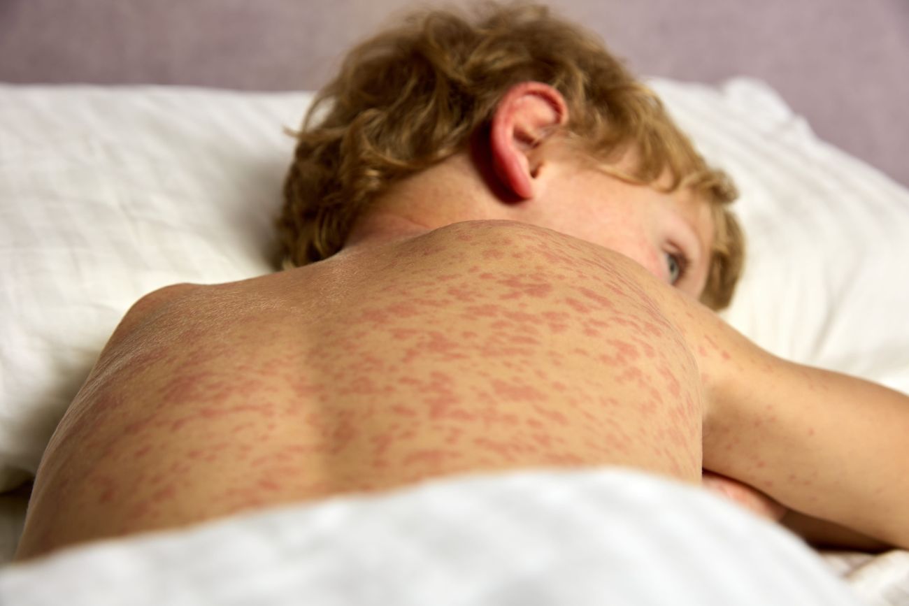
- Viral infections: Many viral illnesses cause temporary lymph node swelling, often accompanied by fever. Infectious mononucleosis (caused by Epstein-Barr virus) is a classic example, characterized by fever, fatigue, sore throat, and widespread lymphadenopathy, particularly in the posterior cervical nodes. Other viruses, such as CMV, adenovirus, influenza, HIV, rubella, and others, can also cause fever accompanied by generalized lymph node enlargement. These are usually accompanied by other viral symptoms, such as respiratory issues, rash, or hepatosplenomegaly, in EBV. Diagnosis can be confirmed through appropriate serologies, such as Monospot or EBV titers, CMV IgM, HIV testing, and others as indicated.
- Kawasaki disease: This is an acute vasculitis affecting young children, characterized by a high fever lasting at least 5 days and multiple mucocutaneous signs. One of the main criteria is cervical lymphadenopathy of ≥1.5 cm, usually on one side. A child with Kawasaki disease typically looks ill, with a persistent fever, conjunctivitis, rash, red cracked lips or “strawberry” tongue, and swollen hands and feet. Recognizing Kawasaki disease is crucial because prompt treatment with IVIG is necessary to prevent coronary aneurysms. Consider Kawasaki disease in any infant or toddler with unexplained fever lasting over 5 days and cervical lymphadenopathy, especially if other clinical signs are present.
- Reactive viral lymphadenitis: In addition to EBV, many common childhood viruses such as adenovirus and enteroviruses can cause fever accompanied by localized lymph node swelling (for example, a young child with fever, runny nose, and a tender cervical node from adenovirus). These cases are usually self-limited; supportive care is enough once more serious causes are ruled out.
- Cat-scratch disease (Bartonella henselae): This zoonotic infection often causes subacute fever and regional lymphadenopathy. A cat scratch or bite typically occurs 1-2 weeks before illness onset. The classic presentation is a child with a tender, enlarged lymph node in the axilla, neck, or groin (where the scratch happened), along with mild fever and fatigue. Bartonella serology can confirm the diagnosis; most cases resolve with azithromycin treatment.
- Tuberculosis and Atypical Mycobacteria: Mycobacterial infections can cause fever and long-lasting lymph node swelling. Mycobacterium tuberculosis usually results in chronic cervical lymphadenitis (traditionally called “scrofula”), often accompanied by general symptoms like low-grade fever, weight loss, or night sweats. Atypical mycobacteria (such as Mycobacterium avium complex) lead to a slow, persistent lymphadenopathy, typically in the neck of children under 5 years old, often without systemic symptoms or only mild fever; the skin over the affected area may appear bluish-purple. If a mycobacterial infection is suspected, perform a tuberculin skin test or IGRA and consider an excisional biopsy for culture. Treatment varies: TB requires multi-drug therapy, while atypical mycobacteria often resolve with surgical excision.
- Systemic JIA (Still’s disease): Systemic juvenile idiopathic arthritis, also known as Still’s disease, is an autoinflammatory condition that can present with daily spiking fevers, transient rash, arthritis, and often enlarged lymph nodes and spleen or liver. It usually occurs in school-age children. Lymph nodes affected by Still’s disease may be moderately enlarged and non-tender. The diagnosis is considered after ruling out infectious and malignant causes of fever, especially when there are additional features like a salmon-pink rash or arthritis. Elevated inflammatory markers and ferritin levels are common, and rheumatology consultation is recommended for confirmation and treatment.
- Lupus or other autoimmune diseases: Active systemic lupus erythematosus (SLE) flares can cause fever, generalized lymphadenopathy, rash, and arthritis, among other features. Lymph nodes in SLE are usually mildly enlarged and soft, indicating reactive hyperplasia. Lupus lymphadenitis can resemble Kikuchi disease histologically, so serologic testing for ANA and other antibodies is useful if SLE is considered in the differential. Dermatomyositis or sarcoidosis (rare in children) are other autoimmune causes that can include lymphadenopathy.
- Hemophagocytic lymphohistiocytosis (HLH): HLH is a life-threatening hyperinflammatory syndrome (primary or secondary) characterized by persistent fever, cytopenias, elevated ferritin levels, and organomegaly. Lymphadenopathy can occur in HLH patients—especially when triggered by infections or lymphoma—but it is not always prominent. Any child with prolonged fever, hepatosplenomegaly, cytopenias, and high ferritin should be evaluated for HLH.
- Malignancies: Certain cancers often present with fever and lymphadenopathy. Lymphomas (such as Hodgkin lymphoma) frequently cause fevers, including Pel-Ebstein cyclic fevers in Hodgkin, night sweats, weight loss, and painless lymph node enlargement. Nodes affected by malignancy are usually firm, non-tender, matted, and progressively enlarge. Leukemia can present with fever, diffuse lymphadenopathy, liver and spleen enlargement, and other signs like pallor or bruising. If unexplained fever and lymphadenopathy persist, especially with abnormal blood counts or other red flags, prompt evaluation for malignancy is necessary (e.g., CBC, LDH, and referral for biopsy). Fortunately, infections far outnumber malignancies as causes of pediatric fevers with adenopathy.
- Kikuchi disease (Kikuchi-Fujimoto disease): This is a rare, benign cause of fever and lymphadenitis often seen in older children and young adults. It typically causes tender cervical lymphadenopathy (often unilateral) accompanied by fever and night sweats. Kikuchi disease is a self-limited condition (resolves over a few months) characterized by necrotizing lymph node histology. It can mimic lymphoma or lupus clinically and pathologically, so diagnosis often requires a lymph node biopsy. There is no specific treatment aside from NSAIDs or corticosteroids in prolonged cases; most recover fully. Consider Kikuchi disease in adolescents with persistent fever and cervical lymph nodes once infections, lymphoma, and lupus have been excluded.
- Periodic Fever Syndromes: Autoinflammatory syndromes can present with episodic fevers and lymphadenopathy. PFAPA syndrome (Periodic Fever, Aphthous Stomatitis, Pharyngitis, Adenitis) is the most common periodic fever syndrome in children, usually beginning before age 5. PFAPA episodes involve high fevers lasting 3-7 days every few weeks, along with cervical lymphadenitis, sore throat, and mouth ulcers. Children are completely well between episodes, and the condition is benign, often resolving spontaneously within a few years. Another autoinflammatory condition, Yao syndrome (formerly called NOD2-associated autoinflammatory disease), can occur in older children or adults with recurrent episodes of fever, rash (dermatitis), arthritis, gastrointestinal symptoms, and sometimes lymph node enlargement. Yao syndrome is quite rare; about 16% of reported cases involve lymphadenopathy during flares. Familial Mediterranean fever (FMF) and TNF-receptor associated periodic syndrome (TRAPS) are other periodic fever syndromes that may sometimes include lymphadenopathy as part of systemic inflammation, although they more commonly feature serositis, rash, or myalgias rather than prominent lymph nodes.
In summary, fever combined with lymphadenopathy in a child should initially suggest infection (bacterial or viral), but inflammatory conditions (Kawasaki, PFAPA, Kikuchi, autoimmune diseases) and malignancies should also be considered if red flags are present. The pattern of fever (acute, recurrent, or chronic) and associated signs are very useful in narrowing down the differential diagnosis. Appropriate laboratory tests (e.g., CBC, inflammatory markers, serologies) should be selected based on the most likely diagnoses suggested by clinical clues.
Lymphadenopathy without Fever: Differential Diagnosis
When a child has swollen lymph nodes without fever or systemic symptoms, the differential diagnosis shifts toward more benign or localized causes. Key considerations for afebrile lymphadenopathy include:
- Recent infection with residual reactive nodes: It is common for children to have persistent reactive lymphadenopathy after an infection, even once fever and illness have resolved. For example, a child who recently had a scalp or throat infection might have an enlarged cervical node that no longer causes pain or fever but remains palpable for weeks. Such reactive nodes are usually soft, mobile, and gradually decrease over 2-6 weeks. As long as the child is otherwise healthy and the node is getting smaller, this condition can be safely observed with reassurance.
- Localized chronic infections: Some infections cause lymphadenopathy with little or no fever. Atypical mycobacterial infection (discussed above) is a classic cause of chronic lymphadenitis without systemic illness – a child under 5 may have a gradually enlarging, non-tender node in the neck (often with overlying skin discoloration) for many weeks, but no fever or malaise. Toxoplasmosis is another infection that may cause subacute lymphadenopathy (often cervical) with mild fatigue but usually no high fever. Cat-scratch disease can sometimes present without fever – a child might simply have an isolated enlarged node in the axilla or neck for a prolonged period after a kitten scratch, and feel otherwise fine. In each of these cases, targeted testing (e.g., Bartonella or Toxoplasma serology, TB testing) can confirm the infection even if fever is absent, and appropriate therapy can be given if needed.
- Malignancy (lymphoma/leukemia): Cancer should be considered in afebrile lymphadenopathy if physical features or the duration are concerning. Lymphomas, especially Hodgkin lymphoma, may present with a painless, firm lymph node (often in the neck or supraclavicular area) without initial fever. The child might otherwise appear well. Over time, additional nodes may enlarge, or other symptoms such as fevers, weight loss, night sweats, or pruritus may develop; however, fever may be absent early on. Leukemia can sometimes present subtly with symptoms such as lymph node swelling, pallor, or fatigue, without accompanying fever. Clues indicating malignancy include node hardness, fixation (matted nodes), abnormal locations (e.g., supraclavicular), accompanying hepatosplenomegaly or mediastinal mass, and lack of reduction over time. Any persistent lymphadenopathy lasting more than approximately 4-6 weeks without a clear cause, especially if larger than 2 cm or accompanied by such warning signs, should prompt further evaluation, including imaging and likely an excisional biopsy.
- Unicentric Castleman disease: Castleman disease (angiofollicular lymph node hyperplasia) is a rare lymphoproliferative disorder that can mimic malignancy. In unicentric Castleman disease (UCD), a single lymph node (or one region) is involved—often in the chest, neck, or abdomen—and the child may have no fever or systemic symptoms. The node enlarges over time and can become quite large. Because UCD is localized, surgical excision is usually curative. Multicentric Castleman disease (MCD), by contrast, presents with generalized lymph nodes and systemic inflammation (fever, weight loss, organomegaly), more similar to a lymphoma-like illness. MCD in children is extremely rare but should be considered if multiple nodes enlarge with inflammatory symptoms and no malignancy is found. Castleman disease requires a lymph node biopsy for diagnosis (showing characteristic lymph node histology), and treatment ranges from surgery (for UCD) to immunosuppressive or anti-IL-6 therapy (for MCD). Although rare, Castleman disease is on the differential for persistent, unexplained lymphadenopathy.
- Rosai-Dorfman disease, also known as sinus histiocytosis with massive lymphadenopathy, is a rare benign disorder characterized by overproduction of immune cells. It most commonly affects children and young adults, and typically presents as large, painless cervical lymphadenopathy, often bilateral, in an otherwise healthy child. The nodes can become very enlarged. Some cases may include mild fever or laboratory signs of inflammation, but many patients lack significant systemic symptoms aside from the enlarged lymph nodes. Diagnosis is confirmed through lymph node biopsy, which shows characteristic S100-positive histiocytes with emperipolesis. Many cases do not require treatment and may resolve spontaneously over time, while those with organ involvement might need steroids or other therapies. In a child with significantly enlarged nodes who appears well and has a biopsy indicating benign histiocytosis, Rosai-Dorfman disease is the most probable diagnosis.
- Kimura disease: This is a rare, chronic inflammatory disorder that causes painless lymph node enlargement in the head and neck, typically in adolescent or young adult males of Asian descent. Kimura disease often features eosinophilia and very high IgE levels. Patients usually do not have a fever but may notice visible swelling, such as in the salivary gland or behind the ear, due to lymph node and soft tissue involvement. It is benign but can be persistent. Although rare, Kimura disease is a possible cause of chronic lymphadenopathy and falls under “miscellaneous” causes. A biopsy is necessary to differentiate it from other conditions, showing lymphoid follicles with eosinophilic infiltrates.
- Sarcoidosis: Although sarcoidosis is uncommon in children, especially in younger ones, it can present with widespread lymphadenopathy without fever, particularly involving hilar lymph nodes seen on chest imaging. It may also cause symptoms like a persistent cough or rash. Any teenager with unexplained intrathoracic adenopathy, possibly along with arthritis or uveitis, could have sarcoidosis. Diagnosis can be confirmed through serum ACE levels and biopsy of a lymph node or affected tissue showing non-caseating granulomas. Sarcoidosis is considered one of the less common causes of lymphadenopathy in older children.
- Chronic benign enlargement: Some children, especially between ages 3 and 10, may have persistently palpable lymph nodes, often in the neck, due to repeated minor infections. This condition is sometimes called persistent generalized lymphadenopathy when no specific cause is identified. If the nodes are less than 1.5-2 cm, soft, and the child is thriving without symptoms, careful observation is appropriate. Additionally, children with eczema or chronic dermatitis on the scalp or face can have persistently enlarged nearby nodes as a result of ongoing skin inflammation.
In cases of afebrile lymphadenopathy, the primary goal is to differentiate between benign, reactive, or inflammatory causes and neoplastic ones. The rate of change (stable or slowly regressing versus progressively enlarging) and characteristics of the lymph nodes are crucial. Many cases of asymptomatic lymphadenopathy in children can be monitored expectantly for a few weeks; for example, viral causes can result in nodes that take 4-6 weeks to resolve. If nodes persist beyond 6 weeks without decreasing in size, or at any point if features suggest a process other than benign reactive, further investigation is necessary.
Rare and Unusual Causes to Consider
In practice, most pediatric lymphadenopathy is caused by common infections or benign reactive hyperplasia. However, for completeness, clinicians should be aware of some rare diseases that can present with lymph node enlargement (often in specialized settings or after common causes have been ruled out):
- Castleman disease: (discussed above) – a rare lymphoproliferative disorder; consider especially if a single large node or conglomerate of nodes persists without diagnosis (UCD), or if multi-node involvement with fever mimics lymphoma (MCD).
- Kikuchi-Fujimoto disease: (Discussed above) – necrotizing cervical lymphadenitis in adolescents and young adults, usually self-resolving; consider it in cases of prolonged fever with negative infectious and malignancy workup.
- Rosai-Dorfman disease: benign sinus histiocytosis with massive lymphadenopathy; consider this when lymph nodes are very large and numerous, but biopsy reveals a non-malignant histiocytic process.
- Kimura disease: chronic head/neck lymphadenopathy with eosinophilia, mainly in Asian patients; a very rare differential diagnosis.
- Autoimmune lymphoproliferative syndrome (ALPS): A rare genetic disorder involving lymphocyte apoptosis that causes chronic non-malignant lymphadenopathy (and splenomegaly) from early childhood, often without severe systemic illness. Children with ALPS have enlarged lymph nodes, autoimmune features (such as low blood cell counts), but no malignancy – specialized testing (e.g., for double negative T-cells) is necessary for diagnosis.
- Immunodeficiency-related lymphadenopathy: Children with chronic immunodeficiencies, such as HIV infection or less commonly chronic granulomatous disease, can experience prolonged lymphadenopathy. HIV, in particular, can cause persistent generalized lymphadenopathy in the early stages, usually without high fever. Always evaluate the child’s risk factors and test for HIV if the history suggests it or if no other cause of generalized nodes is identified.
- Drug reactions: Drug-induced pseudolymphoma or DRESS syndrome (Drug Reaction with Eosinophilia and Systemic Symptoms) can cause lymph node enlargement along with rash and systemic features, but these cases are less common in pediatrics. Serum sickness (e.g., to certain medications) can also cause fever, rash, and lymphadenopathy. A thorough medication history is important to identify these conditions.
These rare diagnoses are uncommon, but pediatricians or pediatric rheumatologists may encounter them in a tertiary care setting. They usually need specialist input and biopsy confirmation. Remembering them helps create a broad differential when common causes are ruled out.
Diagnostic Workup and Management
The method for evaluating lymphadenopathy in children should be systematic and driven by clinical suspicion.
- Watchful waiting versus immediate investigation: If the child appears otherwise healthy, has small (<2 cm), likely reactive nodes (such as after a known infection), a period of observation for a few weeks is appropriate. Many reactive lymph nodes will decrease in size as the immune response subsides. Ensure proper follow-up to confirm that the nodes are improving. Conversely, if there are red flags (such as a very large node, abnormal location, systemic symptoms, or no clear benign cause), do not delay further evaluation.
- Laboratory Tests: Customize labs based on suspected causes. Basic tests often include a CBC with differential (to check for leukemia, cytopenias, and signs of infection or inflammation), inflammatory markers (ESR, CRP, ferritin) if systemic illness is suspected, and LDH and uric acid (which may be elevated in lymphoma or leukemia). If infection seems likely, order specific tests such as Monospot or EBV serologies for mononucleosis, throat culture or rapid strep for pharyngitis, CMV and toxoplasmosis serology, Bartonella henselae serology (for cat-scratch), and PPD/IGRA for tuberculosis. For recurrent fevers or systemic inflammation, consider autoimmune markers (ANA, DS-DNA, etc., for lupus) or genetic testing for periodic fever syndromes as guided by a specialist.
- Imaging: If a node is large or deep, imaging helps characterize it and guides further action. Ultrasound is recommended as the initial imaging modality for children with a neck mass (up to approximately 14 years old). Ultrasound can distinguish solid nodes from abscesses and assess internal features (vascular flow, cystic changes). It is non-invasive and does not involve radiation. For older adolescents or if thoracic or abdominal lymphadenopathy is suspected, a CT scan or MRI may be used. A chest X-ray is a useful screening tool if intrathoracic lymphadenopathy or a mediastinal mass is a concern (e.g., in suspected lymphoma). Imaging is especially important for persistent, unexplained adenopathy to check for additional lymph node enlargement or abscess formation.
- Empiric therapy: In a child who appears ill with signs of acute bacterial lymphadenitis (e.g., a unilateral, enlarged, tender, and red node accompanied by fever), it is reasonable to initiate empiric antibiotics targeting Staphylococcus and Streptococcus while awaiting test results. Response to antibiotics should be monitored within 48-72 hours; lack of improvement might require imaging or drainage. Corticosteroids, however, should be avoided in cases of unexplained lymphadenopathy until a diagnosis is confirmed because steroids can mask symptoms and even temporarily shrink lymphomas, delaying diagnosis. One exception is if a specific inflammatory diagnosis has been made (like Kawasaki disease or PFAPA, where steroids might be used), but steroids should not be used blindly for lymph node reduction.
- When to biopsy: A lymph node biopsy is the definitive diagnostic step when malignancy or unusual disorders are suspected. Indications for biopsy include: unexplained and persistent lymphadenopathy (typically >4-6 weeks without improvement) or progressive cases, nodes that are large (>2-2.5 cm) and not responding to conservative management, supraclavicular or other suspicious locations, and any clinical suspicion of lymphoma or serious illness. In practice, if a node has no clear benign cause and especially if it shows concerning features (hard, matted, fixed, or associated with systemic symptoms), an excisional biopsy is usually recommended. Excisional biopsy (removal of the entire node) is preferred in children because it provides ample tissue architecture for pathology—this yields higher diagnostic accuracy compared to fine-needle aspiration. Fine-needle or core needle biopsy may be considered in certain cases (or for deep nodes), but often can be non-diagnostic in children. Cultures should be performed on biopsy specimens if infection (like mycobacteria) is part of the differential. Remember, excisional biopsy remains the gold standard for diagnosing persistent lymphadenopathy of unknown cause.
- Specialist referral: Consider involving a pediatric hematologist/oncologist if malignancy is a concern or a pediatric infectious disease specialist if unusual infections (such as TB or cat-scratch disease) are suspected and management is complex. A pediatric rheumatology consultation is valuable when an autoimmune or autoinflammatory cause is suspected (e.g., possible systemic JIA, lupus, Kawasaki disease, periodic fever syndromes, or Castleman disease). Multidisciplinary collaboration will help ensure the child receives a timely diagnosis and appropriate treatment.
- Management: Treatment varies based on the cause. Many reactive or viral cases require no specific treatment besides time and possibly NSAIDs for comfort. Bacterial lymphadenitis responds to antibiotics (and drainage if abscessed). Kawasaki disease needs IVIG and aspirin. Autoimmune diseases like SLE or JIA are managed with immunosuppressants. Malignancies are treated with chemotherapy, among other options, after diagnosis. For rare disorders: Castleman disease may require surgery or immunotherapy (e.g., anti-IL6 like tocilizumab for multicentric disease), Kikuchi disease usually only needs supportive care (NSAIDs; short steroids if severe) since it resolves on its own, and Rosai-Dorfman disease might just be observed or treated with steroids if organs are involved.
Conclusion
Lymphadenopathy in children is common, and fortunately most cases are due to benign, self-limited causes (especially infections). A systematic approach—considering the presence or absence of fever, the distribution of nodes, duration, and related clinical features—will guide the differential diagnosis and workup. Always start with a thorough history and physical exam, as they often reveal the likely diagnosis or at least narrow it down significantly. Use targeted tests to confirm the cause, and remember that in a healthy-appearing child, observation can be appropriate. However, stay alert for red flags (large, unusual nodes or systemic symptoms) and investigate those promptly. By evaluating both common causes (like infections, Kawasaki, PFAPA) and less common ones (such as malignancies, Castleman disease, Kikuchi, Yao syndrome, and others), providers can ensure a comprehensive yet sensible assessment. Early detection of serious conditions like cancer or Kawasaki disease can be life-saving, while recognizing benign causes can avoid unnecessary procedures. In summary, evaluating pediatric lymphadenopathy requires balancing caution and prudence—investigate when needed, but avoid overreacting to the many mild, temporary lymph node enlargements that are part of normal immune responses. With a thorough, evidence-based approach, clinicians can confidently manage lymphadenopathy in children and involve specialists when appropriate to provide the best care.
More posts for medical professionals